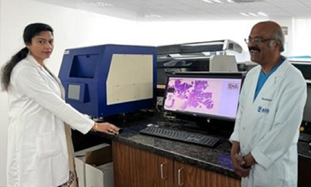
Lumea and OptraSCAN Announce Partnership to Enable Seamless Adoption of Digital Pathology
Lumea, the leader in integrated digital pathology, & OptraSCAN, the top end-to-end digital pathology solution provider, announced a collaboration agreement.
Lumea, a global leader in integrated digital pathology solutions, and OptraSCAN, a leading end-to-end digital pathology solution provider, today announced a strategic commercial collaboration agreement. This agreement brings together Lumea’s comprehensive digital pathology platform with a wide range of digital scanning solutions to enable providers to adopt digital pathology efficiently and cost-effectively.
Lumea combines unique tissue-handling technology with digital technology to create efficiencies for the surgery center, lab, and pathologists, and bring a return on investment to digital pathology.
OptraScan has developed a wide range of digital pathology scanners for small, medium, and large high-volume laboratories with Brightfield, Fluorescence, and Frozen Section for Live View in surgery suites. All products are designed from the ground up to produce high-quality images at a cost-effective price point.
The Lumea-OptraSCAN partnership will enable the following:
Seamless adoption of Lumea’s workflow solutions and AI along with OptraScan’s digital scanners tailored to the needs of a wide range of customers.
Multiple options for leasing, purchasing, or pay-on-demand commercial agreements to find the best financial option for every customer.
A framework for global collaboration with partners to enable better patient management, increased innovation, and ultimately better outcomes for everyone.
“OptraScan set out on a mission to disrupt the digital pathology marketplace by providing high-quality solutions accessible to everyone and reducing the barriers to entry,” said Abhi Gholap, OptraSCAN’s CEO and Co-Founder. “Lumea’s commitment to innovation and quality resonates with the values of OptraSCAN, and we share a common goal to enable wide adoption of digital pathology.”
“Lumea is revolutionizing the way companies adopt and implement digital pathology and we are thrilled to work side by side with OptraSCAN assisting users with the highest quality solutions to improve the ability to read digitally.” said James Thackeray, Lumea Chief Commercialization Officer.
OptraScan and Lumea are also both sponsors of the Digital Diagnostic Summit, a collaborative event where the leaders in digital pathology implementation come together to break down adoption barriers and advance technology throughout the entire pathology workflow. The event will take place September 17-19 in Park City, Utah. Registration is limited to 150 attendees here: digitaldiagnosticsummit.com
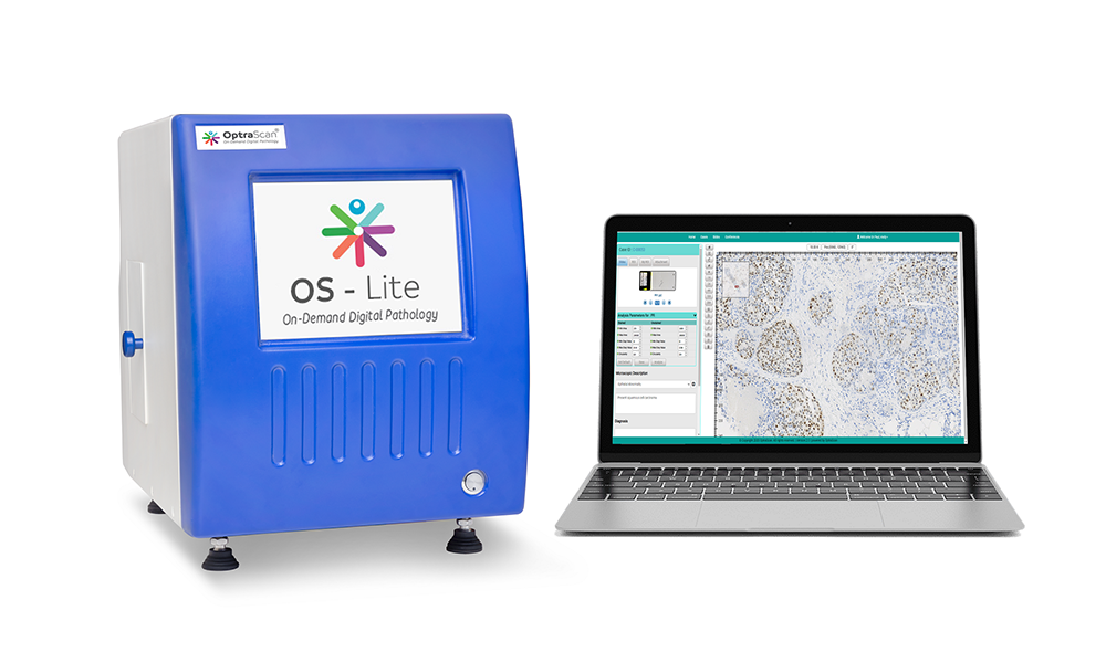
OptraSCAN Launches OnDemand Digital Pathology
Don’t Buy, Just Subscribe.
OptraSCAN®, the leading end-to-end digital pathology solution provider announces the introduction of OnDemand Digital Pathology® solution to meet today’s need for complete digital transformation of pathology laboratories in North America. Laboratories and Hospitals can save hundreds of thousands of dollars spent on Capital purchases by subscribing to OptraSCAN OnDemand Digital Pathology® for a low monthly fee.
OptraSCAN OnDemand Digital Pathology® subscription service will offer OptraSCAN whole slide digital scanners (Brightfield / Fluorescence) with 15-480 slide loading capacity, scanning in about 58 seconds at 40x, a high-end workstation pre-loaded with workflow software, a Telepathology application, and AI analytics packages for IHC / Morphology / Immunofluorescence and Cytology, plus Microsoft Azure Cloud storage and 24×7 support and services.
Speaking at the United States and Canadian Academy of Pathologists (USCAP) Annual Meeting in New Orleans, (Booth #404), Abhi Gholap, Founder and CEO, OptraSCAN said, “Pathology is a cost-conscious market and the current requirement to invest $300-$500k to adopt digital pathology has limited its adoption over the last two decades. With no upfront costs, OptraSCAN scanning devices are compact, easy to install and operate. We ship them to your doorstep and manage them remotely with our 24×7 support infrastructure. We also ensure a 99.99% uptime. With our complete solution to scan, store, share and analyze pathology and cytology images, you can leave your infrastructure worries to us. We will manage, monitor, and deliver to your satisfaction for a low monthly fee. We are getting tremendous market traction with our Year-Over-Year revenues growing at 274% serving the Histopathology and Cytopathology markets.”
About OptraSCAN, Inc.
OptraSCAN has 5 patents on its devices, AI analytics and cloud streaming platforms. OptraSCAN has received CE-IVDR and used at more than 100 sites globally including developing world countries like India, South Africa, and Brazil. Headquartered in California, OptraSCAN has over 23 distributors in USA, UK, Denmark, Italy, Greece, France, Morocco, Russia, China, India, UAE, South Korea, Singapore and Japan.
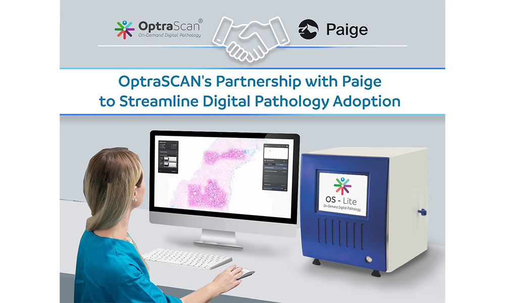
Paige and OptraSCAN Partner to Streamline Digital Pathology Adoption
Paige, a global leader in end-to-end digital pathology solutions and clinical AI applications, and OptraSCAN®, a leading producer of whole slide scanners, today announced a partnership to streamline the adoption of digital pathology technology in the United States, European Union countries and United Kingdom. The companies aim to reduce barriers to digitizing glass pathology slides, which will in turn enhance workflows in pathology and oncology.
Together, the companies will integrate Paige’s clinical AI applications and interoperable enterprise imaging platform with OptraSCAN’s digital pathology scanner to offer advanced end-to-end digital pathology workflow solutions. This provides a streamlined path for pathologists to adopt Paige’s leading suite of digital pathology applications, including the FDA-cleared FullFocus® whole-slide image viewer and AI applications including Paige Prostate Suite and Paige Breast Suite alongside OptraSCAN’s affordable digital pathology scanners. OptraSCAN devices have 15 to 480 slide loading capabilities that digitize the glass slides at 40x in less than one minute per slide with patented composite imaging offering.
“We are excited to work with OptraSCAN to make it easier for labs and healthcare networks around the world to adopt digital pathology workflows,” said Andy Moye, Ph.D., Chief Executive Officer at Paige. “This collaboration is an important step in our mission to unlock the full potential of AI in routine clinical use and provide healthcare professionals with precise insights to help patients get the most effective care.”
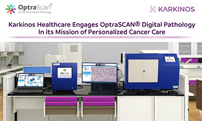
Karkinos Healthcare Engages OptraSCAN® Digital Pathology In its Mission of Personalized Cancer Care
OptraSCAN®, a leading end-to-end digital pathology solution provider, today announced that Karkinos Healthcare has begun using the high-performance OptraSCAN Digital pathology platform to enhance care for their patient population. The capabilities of this digital pathology platform improve accessibility to healthcare and the distributed care model.
The demand for digital pathology has increased as healthcare institutions have struggled to overcome the challenges of the traditional care model of pathologists working in labs with physical microscopes. These workflow problems have been intensified by a shrinking pathologist population at the same time as biopsy rates have increased along with increased cancer screening options. Digital pathology facilitates remote consultations and second opinions to arrive at a diagnosis and initiate cancer treatment more rapidly.
Dr. Ajit Nambiar, Director & Head, Pathology and Lab Medicine at Karkinos Healthcare, explained, “We plan to provide the best possible care to our patients and adoption of OptraSCAN digital pathology solution is an important step in that direction. OptraSCAN is a very capable solution provider, and we are very happy to partner with them.”
The synergies between the two organizations and potential efficiencies were echoed by Mr. Abhi Gholap, Founder & CEO, OptraSCAN, as he stated, “Instant second opinions, remote reporting and permanent digital storage of patient slides are some of the benefits of digital pathology. We eagerly look forward to facilitating them for Karkinos Healthcare. OptraSCAN has seen increasing sales across the world as more organizations adopt their affordable products and services. With 250% growth in our year-to-year global sales revenues, OptraSCAN is a preferred vendor of many small to large laboratories and hospitals worldwide.”
Karkinos Healthcare is pioneering the Distributed Cancer Care Network model in India addressing clinical needs of cancer patients, with a focus on early detection of cancer. The company works with multiple healthcare institutions and professionals in the oncology ecosystem, successfully bringing care closer to patients backed by a robust technology platform. Karkinos already offers its services in the states of Kerala, Tamil Nadu, Karnataka, Andhra Pradesh, Telangana, Maharashtra, Madhya Pradesh, West Bengal, Manipur, Delhi NCR and is planning to ramp up its operations across India. The company is a member of National Cancer Grid, a group of 260+ cancer focused institutions. Karkinos Healthcare is also setting up a Cancer Centre in Jawaharlal Nehru Institute of Medical Sciences (JNIMS) premises at Imphal, in partnership with the Government of Manipur.
About Karkinos Healthcare
Karkinos Healthcare is a technology driven oncology focused managed health care platform for early detection and diagnosis of common cancers. The company espouses the use of a distributed cancer care network working with multiple healthcare institutions and professionals in the ecosystem with a view to get cancer care closer to the persons’ home by decoupling the delivery from the knowledge systems of care.
For more information, visit https://www.karkinos.in/about-us/
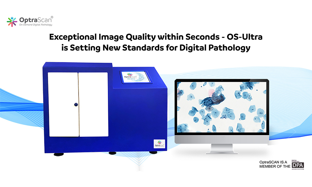
OptraSCAN® receives CE-IVDR for OS-Ultra™ high-performance Digital Pathology System
OptraSCAN® a leading end-to-end digital pathology solution provider has received CE-IVDR marking for the OS-Ultra™ scanners, its high-performance product line of brightfield scanners at an extremely affordable price point, including pay-per-scan based OPEX models.
OS Ultra™ scanners have a loading capacity of 80 to 480 slides. With no-touch continuous loading, it scans tissues or cells with a 15 mm x 15 mm area at 40x magnification in less than 60 seconds. They produce high-resolution images that can be analyzed further with the help of OptraSCAN’s artificial intelligence-based software. This platform is capable of scanning histopathology and cytopathology slides. In addition, it can be configured with 5 to 11 layers for volume scanning or viewed as composite, single layer, fully focused image.
The tray and basket system used by OS-Ultra™ scanners are designed to efficiently transfer and position slides for scanning and prevent slide handling mistakes. For organizations seeking to scale up operations by increasing throughput, OS Ultra™ is an efficient, consistent, and accurate solution.
OptraSCAN’s full digital pathology solutions include scanning devices, image management and viewing software, telepathology software, and image analysis solutions for biomarker and morphometry.
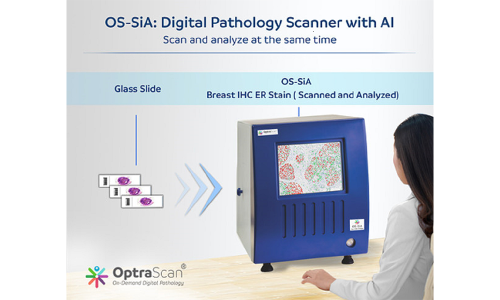
OptraSCAN’s Artificial Intelligence-Equipped Digital Pathology Scanner OS-SiA Granted U.S Patent for Scanning, Indexing & Analyzing of the Tissue Area at the Same Time
OptraSCAN®, the leading end-to-end digital pathology solution provider, announced its digital pathology scanner OS-SiA has been granted US patent – No 2020/0334814 A1 by the United States Patent and Trademark Office. OS-SiA has inbuilt intelligence to scan, index and analyze pathological samples simultaneously. This will benefit the end-user to view the whole slide scanned image along with analyzed output as an overlay during their review process.
The patent describes a technique invented by OptraSCAN, the AI-enabled digital pathology scanner OS-SiA automatically identifies specimens to scan and simultaneously analyzes the tissue or cell area being scanned. OS-SiA is the industry’s first AI-enabled digital scanner that will provide real-time predictive analysis and actionable insights.
Currently, the digital pathology slide scanners are restricted to partial or whole slide image acquisition and digitization into an image. Our next-generation scanner OS-SiA scans and analyzes simultaneously eliminating the need for additional processing applications”. This patent highlights our constant efforts to improve the adoption of digital pathology solutions and support the pathology community.”
OS-SiA can be embedded in the existing series of its cloud-enabled brightfield scanners namely OS-Lite & OS-Ultra. The custom algorithms provide real-time ROI detection while scanning, cell quantification for IHC/HNE markers, and morphological measurements built using the core library. The whole slide image can be viewed in a local / web-based / cloud-based image viewer. The deep learning computational module is provided for self-learning in the scanning device.
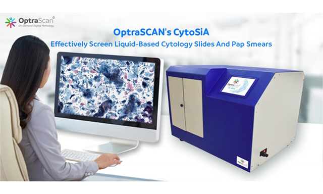
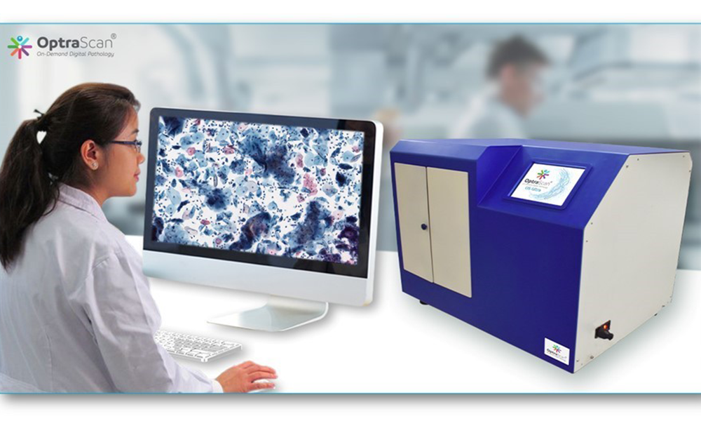
CytoSiA – To Advance Cytology Screening Through Unmatched Image Quality And Artificial Intelligence Based Image Analysis Solution
Cervical cancer is the second most common gynecologic cancer worldwide. This cancer, unlike the others, does not show any symptoms in its early stage, resulting in increased mortality among women. Cervical cancer can be prevented if effective screening measures are set up. Manual analysis of cytology specimens is extremely tedious, time-consuming and prone to human error, thereby generating the need for computer-assisted tools.OptraSCAN’s CytoSiA offers a plethora of features, in order to effectively screen liquid-based cytology slides, and pap smears to differentiate between normal and abnormal cervical cells. In addition to the cells, infections could be further categorized as reactive, endometrial, actinomyces, candida, clue cells, trichomonas vaginalis, and herpes. It also helps in the identification of entities including blood.
The complex multilayer distribution of cells requires multi-layer/multiplanar scanning – a critical component for morphologic analysis for cytopathologists.Conventional scan routines within a narrow focal range has limitations for Cytology smear slides, as cytology slides have three-dimensional cell groups. This complex multilayer distribution of cells requires multi-layer/multiplanar scanning – a critical component for morphologic analysis for cytopathologists. OptraSCAN’s volume scan routine provides a dual mode for scanning and viewing cytology smears; Z – stacking and extended depth focusing algorithm.
Z-Stacking Technology:
A stack of images is generated at different focal planes along the z- axis. The viewing software enables the user to navigate, zoom up and down the different planes to detect the three-dimensional regions in focus.
Extended Depth Of Field:
Volume scan using z-stacks is expensive in terms of scan time and size due to its multilayer nature. OptraSCAN’s extended depth of field algorithm generates a single, entirely focused composite image to effectively, easily and systematically screen an entire image.
Advantages:
Features Of Optrascan’s Volume Scanning Technology:
CytoSiA for rapid yet affordable scanning and analyzing of liquid-based cytology slides and pap-smears. It is a complete solution consisting of OptraSCAN’s digital pathology scanner, storage, and powerful artificial intelligence (AI) algorithms to assist pathologists and cytotechnologists in screening and detection of cervical cancer, pre-cancerous lesions, atypical cells, and all other cytologic categories. Multiple hospitals and pathology laboratories globally have already installed CytoSiA and are witnessing improved patient outcomes, increased efficiency, and productivity needed to cope with the ever-increasing demand of cytology cases.
OptraSCAN Is Disrupting the Digital Pathology Space with Ultra-Fast Scanners
OptraSCAN’s latest addition OS-Ultra modes are designed to assist pathologist to scan images of Immunohistochemistry, HnE and cytology slides in less than 60 seconds.
OS-Ultra is a high-speed whole slide digital pathology scanner that scans tissues/cells of size 15×15 mm area at 40x equivalent in less than 60 seconds. OS-Ultra features a unique no-touch slide loading mechanism to diminish workflow errors and produce high-resolution images. The high-speed scanner comes with solutions – IMAGEPath®, the Image Management System apt for viewing, storing and archiving and TELEPath®, Telepathology software used for real-time, and remote consultations.
OS-Ultra digital pathology scanner is equipped with integrated workflow designed to meet the needs of high-volume laboratories. With OS-Ultra digital pathology scanners pathologists can scan, analyze, store, share and consult with remote pathologists across the globe.
The entire workflow is designed in such a manner that with one click a pathologist or a technician can achieve high resolution image within seconds. OS-Ultra high-speed whole slide scanners is created with the help of pathologists, for pathologists.
OptraSCAN OS-Ultra intends to increase the efficiency and productivity of laboratory workflow.
OptraSCAN solutions are open and scalable that allows pathologists to implement digital pathology solutions without any technical bottlenecks. The solutions can be easily interfaced with various Laboratory Information Systems (LIS).
OS-Ultra technical specifications:
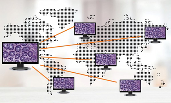
The Need for Telepathology In Developing Countries
Digital Pathology has offered tremendous opportunities to pathologists, be it in the laboratories, universities, or even research centers. It has unlocked new possibilities not only for day-to-day reporting but also for a remote or expert consultation indeveloping economiesor developed economies.
Integration of machine learning and artificial intelligence technologies are assisting pathologists to make an informed decision.
Telepathology: A Connection of Hope
The current circumstances owing to coronavirus outbreak; has triggered a need for remote work on a grand scale. The pathologist community needs a solution that’s quick, cost-effective and will facilitate the transfer of image-rich pathology data between distant locations.
In developing economies there are huge gaps in the healthcare system. Few of them being the availability of expert pathologists for consultation during emergencies; especially in rural areas, and challenges involved in physically transporting the slides.
What is Telepathology?
The use of telecommunications technology to facilitate the transmission of image-rich pathology data between two distant locations for research, consultation, education, and analysis purpose.
To practice telepathology, a pathologist must select the images that need to be analyzed and then make an analysis. A pathologist can study the slides remotely in real-time basis and deliver immediate analysis.
Impact of Telepathology
1. Accessibility of Care
Pathology analysis is critical, concerning patient outcomes. The absence of a local pathologist or absence of expert opinion can harm patient health, create delays and costs, and effect surgery choices and planning.
2. Quality of Care
3. Improved Efficiency
How Can Developing Economies Benefit from Telepathology?
How Is OptraSCAN Assisting the Pathologist Community?
We found three major challenges pathologists face while implementing Telepathology
Here’s How We Are Resolving the Challenges:
1. Cost effective& requires minimum upfront cost- OptraSCAN’s On-Demand TELEPath™ is an affordable, unique automated pathology solution that provides the pathologist community much-needed flexible working model for implementing telepathology with minimum upfront investment.With TELEPath™, one can achieve a considerable increase in working flexibility, accessibility and efficiency to provide better telepathology services.TELEPath™ is a path-breaking solution that can easily bridge the expert opinion gap concerning urban & rural areas.
2. All OptraSCAN solutions can be integrated with the LIS database currently in place at any establishment.
3. Walkaway automation with 24/7 technical support team
On-Demand TELEPathTM Solution Provides
Benefits of On-Demand TELEPathTM:
Telepathology Is Here to Stay
Needless to say, we as a community will experience more such remote logins in future.
Telepathology is already being practiced to diverging degrees worldwide. Taking advantage of digital solutions laboratories are evolving rapidly and there is an immense opportunity for the field to expand further and unlock complex medical puzzles.
As digital pathology enables collaboration between pathologists as well as within cross-disciplinary teams, pathology labs will be able to offer expertise to regions beyond their current scope where experienced or specialized pathologists are scarce.
Pathologists now need to embrace and take advantage of what these new tools offer in adding objectivity to one’s approach.
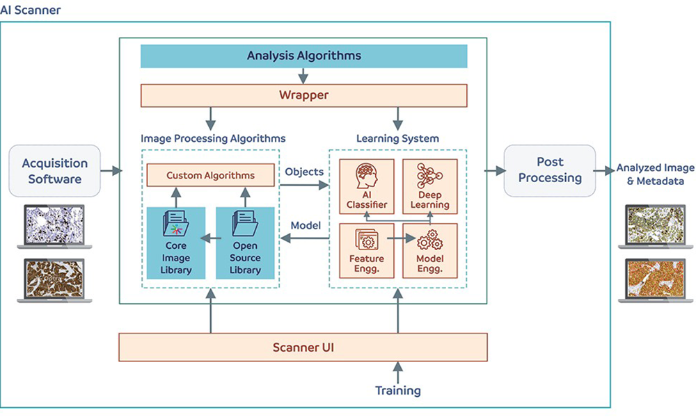
AI-enabled scanner: an example of the machine learning paradigm of feature learning
It is universally known that Digital Pathology (DP) is the process by which glass slides are digitized to produce high-resolution images. However, the promise of digital pathology and the paradigm shift from microscopy to digitization is not merely transfer of an image from glass slide to a monitor; or to share, modify or analyze the image but rather the potential to transform the digital image by augmenting the human eye with information/data to perform intelligent interpretations (algorithmic approaches) that cannot be gleaned by the pathologist’s eye. These digitized slides afford the possibility of applying image analysis techniques to DP for applications in detection, segmentation, and classification. In short, the next breakthrough innovation in digital pathology could be a next-gen smart scanner, which is driven by artificial intelligence and deep learning concept specially designed for the ease of pathologists to scan, index and analyze the tissue slides – all at the same time.
Algorithmic approaches driven by AI and ML technology, have shown to be beneficial in many contexts as they have the capacity to not only significantly reduce the laborious and tedious nature of providing accurate quantifications, but to act as a second reader helping to reduce inter-reader variability among pathologists. Thus, it aids the pathologist to not only look at many thousand cells but pixels which will enable him/her to perform precise measurements of histological patterns on a whole slide image in his/her precision medicine arsenal. “On the fly image analysis solution” embedded in AI-enabled digital pathology scanner is one such algorithmic approach; a combination of artificial intelligence (AI) and Machine Learning (ML) technology.
Characteristics of smart scanner:
A scanner with AI & ML based technology provides fast and accurate diagnostics support in improving medical research.
Advantage of smart scanner:
These systems offer further advantages by integrating the workflow into the operational environment that is hardware agnostic and lets third parties’ plugins thereby making it a good fit. Augmentation that automatically counts cell types enhances standardization and reproducibility will complement the work of a pathologist by providing more accurate and reproducible analysis of key factors eventually improving patient outcomes.
However, the question bothering the pathology community remains as is! As the technology matures, will computer algorithms outperform human pathologists in both accuracy as well as speed and go beyond assistance, replacing human pathologists eventually? The answer is NO. Artificial intelligence (AI) as a “powerful tool” uses the accuracy of machine learning for complex scoring as well as computational tasks and the cognitive power of the pathologist to analyze complex tissue architecture, each performing to their own strengths. Once in the digital realm, artificial intelligence and deep learning technologies in combination with computational pathology techniques with the humongous data generated will help train deep convolutional neural networks which will mimic the human cognitive capabilities. Multilayered convolutional networks will then perform all complex computational tasks and reveal more information to answer questions about a given disease.
Thus, digital pathology will bring this positive change by replacing the microscope, not the pathologists.
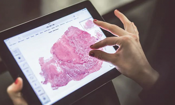
Digital Lab-as-a-Service – a convenient way to adopt digital pathology systems
Many healthcare organizations across the globe today are shifting towards digitizing the age-old practice of scanning and viewing glass slides through microscopes. They have realized the potential of digital pathology and digitization of slides that has a direct impact in productivity and efficiency gain. There’s no doubt that digital pathology is bringing a paradigm shift in the way we look at pathology. It is widely regarded for providing accurate cancer diagnosis and bringing the pathology community closer.
However, many organizations are compelled to rely on the conventional microscopy. This could be due to various economic and social factors, ranging from financial constraints to limited internal resources to deploying and managing the system itself!
Although the above-mentioned reasons are inevitable, they should not deter an organization or the pathologists from adopting digital pathology. There are many ways organizations can implement digital scanners as well as avail easy scanning of whole tissue slides in their eco-system, without spending a single buck, upfront!
Digital Lab-as-a-Service (DLaaS) solution:
Digital Lab-as-a-Service is one-of-a-kind solution; wherein Digital Pathology Systems help the pathology labs, hospitals or any healthcare providers to take advantage of digital pathology benefits. Digital Pathology Systems provider will set up a digital infrastructure including whole slide scanners, software, storage servers, etc. within the hospital or lab. Customers will thus pay per scanned slide towards operational expenses post scanning of slides.
Economical Digital pathology scanner offers numerous benefits, such as
Who all can avail Digital Lab-as-a-Service (DLaaS) solution?
Any healthcare organization or healthcare provider can avail the DLaaS, irrespective of their economical condition. However, at a broader level, following could be the benefitting organizations:
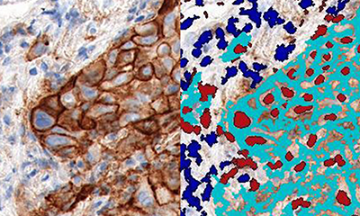
OptraSCAN immune-oncology solution for Programmed Death Ligand 1 expression on tumor and immune cells
The field of immune-oncology biomarker is exploding dramatically. Immunotherapy agents overcome tumor evasion through targeted immune checkpoint inhibitors and scientifically driven combination therapies including multiple immune-stimulating and chemotherapeutic agents. However, not all patients are benefited from immunotherapy and precision medicine requires tremendous understanding of the tumor microenvironment. Complex tissue architecture, heterogeneous biomarker expression as well as visual and cognitive traps affects visual interpretation by pathologists. Moreover, increased requirement of precise diagnostic cut-points of the biomarker measurements poses a problem for pathologists, who are asked to scan thousands of cells in a tissue section and to arrive at a score accurately in order to give a result that can predict a response to the targeted therapy. The promise of digital pathology and image analysis tools help overcome these challenges by performing intelligent interpretations that cannot be gleaned by the pathologist’s eye.
Immuno-Oncology image analysis solutions
Flagship Biosciences developed a platform for understanding the abundance and spatial relationships of different immune cell types and for immune cell clustering using duplex immunohistochemistry. Researchers also adopted HALO platform by Indica labs for digital quantification of PD-L1 tumor cells in melanoma. Another area of concern is the availability of multiple PD-L1 diagnostic immunohistochemistry (IHC) assay and lack of clarity on immunolabelling parameters determined by pathologists and the comparability across assays. A customized Image Analysis scoring PD-L1 scoring solution to permit a quantitative comparison of the 4 PDL1 IHC assays was developed by Definiens that enables to quantify the percentage of positive tumor cells on a whole slide image for 4 PD-L1 assays by co-registration of images and comparing identical areas per case. Aperio Image Toolbox and GENIE image analysis tool (Leica Microsystems) quantifies membrane PD-L1 expression in malignant epithelial cells and macrophages based on Region of Interest analysis.
Optra PD-L1 automated whole slide scoring for tumor and immune cells
Biomarker expression in lymphoid or other immune effector cells is a special challenge for pathologists to measure immune phenotypes, which may predict patient responses to immunomodulating therapies, was observed in the Blueprint 2 study. To overcome the aforementioned challenges, Optra PD-L1 automated image analysis approach generates separate scores for interpretation of PD-L1 IHC expression on both tumor and immune cells without the need of multiple biomarkers for immune cells in the same tissue section in a single read. While all the features/functions are available for integrating, analyzing and collaborating on a single platform; OptraSCAN and image analysis suite can support whole slide scanning, integrated image analysis, image registration and quantification of tumor and/or immune cells based on biomarker content to aid immune-oncology development.
Role of digital pathology in improving healthcare in developing nations
Penetration of digital pathology in North America and Europe in research centers, laboratories, and universities is on rise in the recent years. Digital pathology is predicted to follow the successful path of radiology in near future in terms of acceptance. It has been possible due to obvious advantages over conventional microscopy. The possibility to scan and store the entire tissue slide in digital formats has opened many avenues. The sharing of digital image between pathologists for second opinion or consultation has become easier, faster, and safer. Improvement in communication bandwidth and storage facilities has made Telepathology possible for peer reviews. Overall, pathology workflow has become efficient because of the digitization process. Image processing technology in combination with machine learning and artificial intelligence has revolutionized digital pathology in both qualitative and quantitative assessments.
Challenges in digital pathology adoption:
However, with so many advantages, there is still some reluctancy in adopting digital pathology in both developed as well as emergent nations and there are several factors accounting for the same. The ratio of pathologists to population is already low in developed countries and is much lower in developing countries. Due to this huge gap, a pathologist is overloaded with slides that needs to be scrutinized. The mental fatigue affects the efficiency and accuracy of the pathologist. There are other issues apart from manpower deficiency, such as lack of infrastructure, economic constraints, poor quality control in slide preparation and handling, absence of support systems like LIS to manage the workflow, and a large population that hampers the overall quality of healthcare. There is an apparent increase in cases of non-communicable diseases, including cancer, diabetes, etc., in developing nations that require accurate pathologic diagnosis.
How digital pathology can overcome these challenges?
In rural areas, the absence of trained professionals necessitates expert diagnostic consultation from urban centers equipped with better infrastructure. Movement of physical slides, patients, or professionals for consultation is time consuming and an expensive proposition. This also increases the workload on few professionals in urban settings. Digital pathology can help bridge this gap. Digital Pathology Scanner that offer very competitive scan times and an affordable price tag are the need of the hour in emergent nations. The AI/ML-based quantifying solutions built with digital scanners will complement the pathologist in accurate quantitative analysis of slides. Innovative low-cost solutions like on-demand pathology systems, where the user pays per slide and not for the whole package, are being explored by some pioneers in this field. The IT revolution is already pacing in developing countries, with the advent of smart phones and tablets usage increasing with each generation in both the urban and rural areas. There is tremendous scope of growth for telepathology due to rapid advances in information and technology. Primary diagnosis and training in remote areas, secondary opinions, and referrals could be few of the applications that can be very well addressed by Telepathology effectively.
Role of digital pathology in education & research:
Digital pathology could also be helpful in developing countries in medical schools and universities. The system can improve the training process of technical staff and future pathologists. The digitized slide can be viewed multiple times by as many numbers of students at once. The difficulties like the deterioration of slide quality in terms of stain fading and efforts in maintenance of slides can be removed with easy adoption. The cloud-based systems, where the students and teachers can view the virtual slides will make learning and understanding easier as students and teachers will have access to specialized services like IHC, CISH and FISH, which are otherwise not commonly available in remote areas. With improvements in basic infrastructure like information technology, reliable electrical power, and investment in health care programs, digital pathology can bring myriad changes in improving the healthcare services in emergent nations. In my view, an expert pathologist located at a central clinical setup, receiving virtual slides taken across remote areas (probably through affordable scanners) for his review, would be an ideal healthcare model in developing countries.
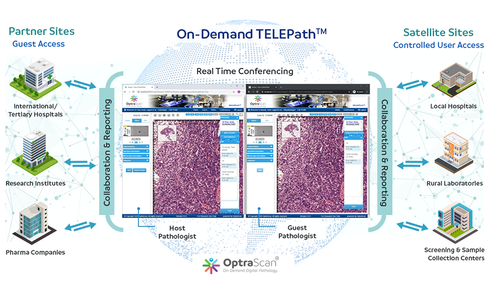
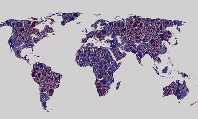
Global TELEPath® Network
Technological innovations continually nurture the process of globalization and the way we deliver healthcare. Telepathology is one such change of delivering remote pathology services and a value-added clinical benefit for better patient care. With its direct effect on healthcare industry, this new paradigm of international telepathology will facilitate cross border collaborations and access to pathology services around the world. Apart from its benefit to the underserved and rural areas in recent years, international teleconsultation has grown and provided a novel source of revenue to pathologists and vendors.
A plethora of such telepathology platforms have been undertaken to serve a multitude of users across the world. One of the earliest was been developed by University of Basel with their global network enabling examination of over 15,000 telepathology cases where static images was the mainstay of image exchange. Virtual International Pathology Network, several telepathology networks established by large academic institutions like University Health Network in Canada, UPMC, MD Anderson in Texas and the Massachusetts General Hospital in Boston are the more recently established providers of expert consultations with adoption of Whole Slide Imaging (WSI). Many commercial vendors with their business partners practicing international telepathology have also started offering telepathology portables e.g Aperio ePathAccess, DP3 platform. Corista, ProNet. XIFIN are a few leading ones. These contemporary telepathology platforms who have adopted WSI enable remote viewing in one of the two ways: either by remote sharing or uploading of images for expert consultations. The former requires strong cooperation between all sectors of the service providers and the latter may suffer delays due to image transmission. Also, these dynamic telepathology platforms are more expensive, and mandate the use of proprietary hardware/software at the receiving and sending end. All the aforementioned challenges hinder interoperability.
Taking advantage of the shift in technology over the past decade, we at Optra have simplified the mode of delivery and have developed an open access platform, TELEPath® with secure, user based logins which supports secure data transfer, patient confidentiality and data encryption accessible even on mobile cellular devices. This hybrid system combines the elements of static, dynamic and real time collaborations which is hardware agnostic without the need for large digital files and image viewer compatibility for different file formats including Z stacked images for interpreting cytology teleconsultations. Key important factors for delivering quality care across borders like quality assurance measures, monitoring timeliness of reporting with a provision for pertinent clinical information, sustainability, flexibility and a bidirectional communication between parties are the currently offered practical solutions to support these collaborations considering the needs of all key stakeholders.
With the draft bill currently before the US Congress to facilitate telemedicine, the practice of international telepathology has a promise to show a positive impact on global patient care.
Intelligent Digital Pathology
Over the past few years, deep learning, artificial intelligence (AI) and healthcare AI has become a hot topic owing to development of advanced computer-assisted analytical tools. Pathologists too have embraced this revolution with the recent FDA approval of whole slide scanners. The promise of digital pathology is not merely transfer of analog data from glass slide to a monitor but augment the human eye with information/data to perform intelligent interpretations. AI and deep learning technologies mimic human cognitive capabilities using sophisticated imaging processing techniques to train deep convolutional neural networks. These trained multilayered convolutional networks then perform all complex computational tasks and reveal more information answering queries pertaining to a given disease.
AI/deep learning systems offer further advantages by integrating the workflow into the operational environment that is hardware agnostic, thereby making it a good fit. A larger set of variables can thus be assessed and analyzed by these computer algorithms on the digitized tissue sections. The inevitability of these digital transition AI tools will be particularly beneficial as the industry moves towards value-based payments. Experts in this field suggest that healthcare providers who pay for performance will appreciate the speed and predictive power of AI. Pathology specialty, as prompted by some, will soon metamorphose into a role of “information specialists” to make room for new technologies.
Image analysis algorithms are already being used for automated or semi-automated immunohistochemistry (IHC) quantification, thereby facilitating standardization, speed of analysis and objective reporting to complement the role of the pathologist. Thus, an early intervention towards making AI as an “assisting tool” is combining the human cognitive capabilities and the predictive power of computational algorithms for providing insights into patient specific treatment options.
Intelligent digital pathology will thus bring about a paradigm shift from conventional microscopy to pathology AI.